


This disease is poorly examined, although several thousand observations with diagnosis and subsequent treatment have been described.
A lot of diversity and non -specificity of the clinical image of varicose veins of the pelvis lead to gross mistakes from the diagnosis, which influences the consequences in the future.
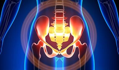
Characterization of varicose veins of a small pelvis
The veins of the pelvis are several times longer than the arteries, which determines their great capacity.This is due to the phylogenesis of the pelvic region's vascular system.The veins of the pelvis have high adaptability and may be predisposed to restructuring, which contributes to the formation of a densely interwoven network.
The speed and direction of the blood flow is regulated by valves that are controlled by complex humoral mechanisms.The valves expose the pressure in different parts of the venous network.
When the valves stop performing their functions, the stagnation of blood develops, this leads to the pathology of blood vessels and the formation of varicose veins.The uniqueness of the veins of the small pelvis lies in the fact that the vast ligaments of the uterus, which can limit the lumen of the ship, which can limit extensive, and cause pathology.
Causes
The pathological dilation of the veins of the pelvis can be due to the following reasons:
- Violation of blood drainage pathways;
- Venous barrel prison;
- Compression of collateral tribes due to the changed position of the uterus, for example in retroflexia;
- Valve -egg failure (congenital or acquired);
- Obstructive Post -Phlebital syndrome;
- Pathology of the connective tissue;
- Arteriovenous angiodisplasia;
- Sitting longer, hard physical work;
- Varicose veins of the lower extremities;
- Pregnancy (3 or more) and birth (2 or more);
- Diseases of the female genital area (chronic salpingoophorite, ovarian tumors, uterine myoma and genital endometriosis);
- The adhesive process of the pelvic organs;
- Obesity.
Classification according to degree of disease
In terms of the extended vein, the following degrees are differentiated:
- Up to 0.5 cm, "corkscrew" vascular stroke;
- 0.6-1 cm;
- More than 1 cm.
Options for the course of the disease
- Varicose veins of the perineum and vagina;
- Venous thoroughbred syndrome of the pelvis;
Symptoms
- The most common - most common pain in the lower abdomen, perineum after long static and dynamic overlay.The pain increases in the second phase of the cycle after hypothermia, tiredness, stress and deterioration of various diseases.
- The feeling is "not comfortable", pain with sex and afterwards.
- Dysmenorrhea - menstrual disorders, including pain syndrome.
- Secretation, more than normal, galls of the genital tract.
- The stagnation of blood leads to infertility, inability and termination of pregnancy.
- Violations of urination as a result of the expansion of the veins of the bladder.
Diagnosis
The diagnosis of the disease is only successful in only 10 % of cases.
The palpation of the inner walls of the pelvis makes it possible to feel elongated seals and venous notes.When examining in the mirrors, the cyanosis of the mucosa of the vagina is visible.
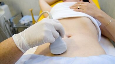
The choice of selection is an ultrasound study with colored mapping, with which you can not only identify seizures of ovarian veins, but also the venous thrombosis, post -trombic occlusion.With ultrasound it is visible, "worms", the structure without reflection of the signal, located on the side surface of the uterus.
The effect of the Doppler study is based on "tinting" in blue and red color, venous and arterial blood.
The apparatus for the ultrasound examination using a special program recognizes the blood movement from the sensor and calculates the blood flow speed and the type of vessel in the other direction.
However, the exact definition of the vein is or the artery remains behind the doctor.The Doppler method works in almost all cases, our body dictates exceptions from the rules, since blood that flows from the heart is not always arterial and vice versa.
An ultrasound diagnostic doctor sees an arterial or venous ship, its size, blood flow rate and many indicators that an ordinary person does not need, but an important role in the diagnosis.Use transabdominal and transvaginal sensors.
In 5.7% of cases, the disease is recognized by accident during screening.Usually the diameter of the Vena -OVARIC is 0.4 cm.
CT and MRI have great accuracy.With these methods you will find accumulation of varicose veins in tapes of the uterus, the ovaries and these organs.It is possible to determine the simultaneous pathology.
A very reliable method is a phlebographical study.
The contrast is carried out in the amount of the Valsalva test against blood flow.In this way you can see valve errors.
Retronoscopy on the left, kidney phlebography, super -sequenic phlebovarioskopy and phlebovariography on both sides are also used.With these methods you can determine the hemodnam and anatomical changes in the kidney veins and the places of falling in Gonadny veins.
Super fever -phlebovarioscopy is carried out by catheterization of gonaden veins by a counterattot dead femur or subclavia venue with the subsequent introduction of the contrast.
Most blood from the plexicosis polled plexus is thrown away by the ovarian vein.However, under high blood pressure conditions, it appears through non -confectioned uterine veins into the inner Iliacal vein.The plexus of the veins through which the drainage can occur includes sacred and plexus of the bladder.
In the left phlebo -icigography, 3 stagnation stages in the Cluster Plexus of the left Egg Day are distinguished:
- There is no discharge from the plexus of the left ovary or it is carried out on an additional short path.
- There is an additional long way.
- Two additional discharge routes or another and one additional aid are visible.
At 2 and 3 stadiums, varicose veins of the Cluster Plexus of the right ovary are formed.
Laparoscopy is used for differential diagnosis.Pathologically confused veins are in the ovary, in the direction of the round and wide ribbons.They look like large cyanotic conglomerates with a thin and tense wall.
The complexity of the diagnosis lies in the fact that the disease is often hidden behind the signs of the inflammatory process by clinical manifestations, which are masked under endometriosis, prolapse of internal organs, postoperative neuropathies and many extingenous diseases.
Treatment
The main goal of the treatment is to remove reflux in the veins.Conservative treatment is used in the early stages of the disease.In the late stages of the disease, the treatment of choice is an operation.
Conservative treatment

It is to normalize the tone of veins, an improvement in hemodynamics and trophic processes.
Symptomatic treatment for the elimination of individual symptoms.Non -steroidal anti -inflammatory pain in the event of bleeding - hemostatic therapy.
The most important drugs in conservative treatment are venotonic drugs and anti -plattelettes.
Phlebotonics - improve the tone of the vessel wall and improve blood flow.With this disease, it is better to consult a gynecologist about certain medication.
Physiotherapy exercises are an important method.
Surgical treatment
- Revival varicose veins.
- Gonado-Cavalry Shunting.
- Sclerosis for laparoscopy.
- Occrames of ovary veins using X-ray endovascular methods.
Folk remedy
Since the main thing for the occurrence of the disease is the weakness of the flap apparatus, all folk remedies are used for the varicose expansion of the veins of the lower extremities for this pathology.
Those used most frequently: ordinary hazelnut, hops, nettles, horse chestnuts, dandelion root, tea mushroom, willow, oak, St. John's wort, a series, flower pollen and many other plants.
It is effective: treatment with oak baths, chestnuts, pastures, chamomile, pharmacy, drying, St. Johns Würze.
prevention
- The first thing to do when complaints, predictors or illnesses are listed at the top - contact a gynecologist.
- It is necessary to normalize the work regime and to rest.Try not to stay in an upright position for a long time.
- Perform exercises for the prevention "pedal", "stand-up stack", "foot legs"
- Stick to the diet: Eat foods with a high content of vitamins E, R, C, try to eat only white meat, less fat, to replace it with fruit, vegetables, cereals.
- Drink a sufficient amount of liquid, but no less than 1.5 liters per day.
- Get rid of bad habits of excess weight.
- Contact the doctor present by wearing compression lines.It will improve blood drainage from the lower extremities, which means that there is less stagnation in the pool.
- Avoid bathrooms, saunas, steam trees, hot baths.
In order not to get sick with such a difficult disease, the preventive recommendations listed above must be observed.Treat your health as the most valuable in life.
With the slightest suspicious symptoms that you cannot get rid of several days, you should consult a doctor.He has to provide them with highly qualified help and save them from suffering.